CHAT

Wednesday, October 19, 2011

Helping Hands New Procedures Revolutionize Treatment of Dupuytren’s Disease

 BY DAN CHASE

July 2011

From left, Drs. Jeffrey Wint, Bruce Wintman, and Richard Martin at the Hand Center of Western Massachusetts.
Helping Hands
New Procedures Revolutionize Treatment of Dupuytren’s Disease
 BY DAN CHASE

It has been called many names, from the ‘Viking Disease,’ due to its prevalence among people of Northern European ancestry, to palmar fibromatosis. But to most patients and doctors, it is named for the French surgeon Baron Guillaume Dupuytren, who described an early treatment for the condition.
Dupuytren’s disease is a contraction of the fingers due to abnormal thickening of tissue. At the Hand Center of Western Massachusetts in Springfield, Drs. Richard Martin, Jeffrey Wint, and Bruce Wintman told HCN that the treatment of the disease has just been given a big helping hand. Two of them, in fact, in the form of new procedures that do nothing short of transform the way patients are given relief.
To get a better understanding of the mysteries of Dupuytren’s, which doesn’t have any specifically known causes — only a series of predispositions in patients — Wintman described the symptoms.
“There are different types of tissue in the hand,” he explained. “There’s skin, muscle, bone, and also a tissue that strands throughout the soft parts to give it form, called the fascia. In Dupuytren’s, your body makes a little bit too much of it, and an abnormal type of it at that.
“As a result,” he continued, “you start to get nodules in the palm into the fingers, and sometimes these can coalesce into a cord of tissue that can actually curl the finger down. That cord can be so thick that you can’t actually straighten it out on your own, even though the joints are normal and tendons are working. That abnormal fascial cord is flexing the fingers down. And that’s what patients complain about, that inability to straighten out those fingers.”
There is no pain particular to the abnormal fascia, but the lack of mobility can lead to problems with work and quality of life.
“It can become so cumbersome, and for some people can become so severe,” Wint added, “that we’ve had some patients say, ‘if you can’t do anything for it, cut the hand off. I just can’t do anything with it.’ That’s the extreme, though. Here, we tend to see anything from simple nodules to cords.”
Historically, the treatment for Dupuytren’s involved an open surgical procedure to remove the abnormal tissue, a process which also involves several months of physical therapy to restore mobility in the hand. However, new developments in the treatment of this condition have severed that downtime to the extent that a patient can have one of two new procedures — done in the office and not the surgical theater — and have immediate results with little aftereffect.
“In some cases,” Wintman said, “a patient can have one of these procedures during the week and be golfing by the weekend.”
This month, HCN sat down with the doctors of the Hand Center to find out what these new processes are, and how they are giving an important helping hand to the treatment of a life-altering condition.
General Contractor
Dupuytren’s is, as mentioned, a bit of a mystery, for there are no proven causes. But doctors say there are certain risk factors, including geographic ethnicity, age (over 40), a history of alcohol use, and, perhaps most importantly, those with a family history of the condition. While the disease is most commonly seen in men, it is not uncommon for women to contract it.
When the first signs of Dupuytren’s are noticed, the tendency is not to take immediate action, Martin said.
“If you get a little nodule, a little bump, there’s not a lot that needs to be done,” he explained. “However, when that becomes a cord, that’s the time when most people feel the need to do something about it. In years past, traditionally we would wait on treatment until it became a significant problem for the individual.”
Wintman said the “flat-table test” is a point of entry for his patients. “It’s usually not a pain situation whereby someone will want to seek treatment, but rather a lifestyle condition,” he explained. “If you can’t lay your hand flat on the table, then you may wish to proceed with treatment.”
For many years, what happened next was a pretty straightforward surgical process. If the nodule or cord was not contracting the fingers, nothing would be done, but the affected areas were kept under observation to monitor any marked development. If the progress contracted the fingers significantly, the hand would be incised, and the abnormal tissue cord removed. Wint said that this was a straightforward surgical procedure.
“The problem isn’t taking that cord out,” he explained. “That’s fairly easy to do. After an hour of surgery you can remove it. The problem is what happens to the hand afterwards. If it’s used to being bent, then the skin may be inefficient — it’s used to being pulled down. The tendons may not be springy right away, and the joints may want to reassume that bent position. So, someone who has a simple surgical removal to get rid of that cord needs to now go into a splint, sometimes full-time, up to a month, and into rehabilitation for three to six months after.”
Recurrence rates are a fact of life for patients with Dupuytren’s, and even with full removal of the contracting cord, the abnormal tissue can return in several years’ time. Also, if the original surgery involved the removal of a nodule, there’s a risk of not excising enough tissue, which Wint said could cause the abnormal tissue to return with increased vigor.
Handle with Care
While open surgery had been the only means for patients suffering this condition to return functionality to their fingers and hands, science has come to their rescue.
Two new procedures, as differentiated from surgery since they can be done in physicians’ offices, have come to the market within the last five years. One of these new techniques, a collagenase injection, was made available only one year ago.
That drug is called Xiaflex, and Wintman explained the process.
“You give a small shot of this enzyme in one area in the cord, and it eats through the collagen which the Dupuytren’s fascia is made from,” he said. “So it dissolves through the collagen, and then disrupts the cord at that one level. You come back the next day for manipulation, and the finger can go straight at that level after the manipulation.”
The second process is called needle aponeurotomy (NA), and essentially is a micro-perforation of the cord.
“We take a needle and actually make a series of puncture wounds along the length of the cord that’s keeping the finger bent down,” Martin said. “This breaks up the cord at various levels using the needle, almost like a tiny scalpel. The cord then actually breaks apart at these different levels. And then, the finger can straighten out. This doesn’t remove the tissue; you’re just breaking it up.
“As a result,” he continued, “afterward there are just a series of exterior puncture wounds that need to heal, typically over the course of five to seven days. Patients go home with some Band-Aids and an ice pack, and they’re moving and using their hand right away. Typically they don’t need a splint, although we might give them one to reinforce the gains we’ve made. But that is rare.”
Choosing between the new procedures comes down, ultimately, to dollars and sense: often an insurance carrier might cover one but not the other.
Also, the NA can involve several affected areas in the hand on one visit, whereas the Xiaflex must be administered only in one area every 30 days.
“So if someone comes in with three fingers affected, that’s a bit of a stretch,” Wintman said. “But some people want to try the medication over the four months.”
However, he added, “a patient might have co-morbidities. They might be on a blood thinner, and you wouldn’t want to give them an injection of an enzyme that eats tissue, because there would be too much swelling. Some people may have skin issues and not want to be exposed.”
But it can come down to a patient’s word-of-mouth referral to determine one over the other, Wint said.
Around the Bend
Sadly, science has not cured Dupuytren’s once and for all. The same recurrence rates for the abnormal tissues does still apply to these two new procedures, and, in fact, the time until reappearance of nodules and cords is often just a few years’ time. But, doctors agree, the ease with which both Xiaflex and NA can be administered should allay any fears of a return visit.
But has science sounded the death knell for the surgical open procedure?
“Well, it does reduce that frequency significantly,” Wint said. “Since that used to be the only solution, it was that or nothing. I had some people who, because of their work, really couldn’t take three to four months off. And you had to try to prepare people, also, because it could have even been longer.”
Martin said there are patients who still say, “I want it out,” and that does mean surgery. And for a patient with a history of severe Dupuytren’s, or situations involving prior complex surgeries, the new procedures might not offer any assistance.
“Their anatomy might be markedly distorted; they may have had skin grafts done elsewhere,” Wintman said. “Also, you can have contractures of the joints — the joint itself is scarred in. It’s not that you can feel a clearly defined cord anymore, but there might be scar tissue that keeps the fingers contracted. And, unfortunately, you really can’t do anything with that in the new procedures.”
But one thing the two new techniques will hopefully bring about is a future whereby doctors don’t see such severe cases of Dupuytren’s. “As more and more people read about these procedures, or hear about them from friends, they’ll realize that the need for the surgery will be less and less,” Martin said.
His colleagues nodded in agreement when Wintman said, “it’s incredibly satisfying for us, because when the patient leaves the office, they will have noticeable results. They walk in with a deficit, and they leave here with straight fingers.”

Why does some carpal tunnel numbness and tingling go away for many people after surgery, but for some symptoms persist?


Will my sensation come back or be normal after surgery?

While the goal of carpal tunnel surgery is to relieve the pressure on the nerve not everyone will respond the same to surgery

Some patients will have immediate return of sensation while some will take longer. Some will notice an improvement right away but still feel tingling and will describe this as "numb" The return of sensation is dependent on many factors including age, general health, duration of symptoms, circulation and the actual mechanical severity of compression. This video shows some drawings that may help to explain why.

In very severe cases while decompressing the nerve stops the carpal tunnel syndrome from getting worse, full recovery of sensation may not be possible. Often this is seen in patients who have muscle wasting noted prior to surgery and in those with longstanding complete numbness and elevated two-point discrimination. Of course there are many in these categories that improve despite having very severe cases.

Having a severe case where you are not sure if you'd have full recovery is not a reason to put off surgery, as progression is likely if nothing is done.

How about my strength?

This is a very difficult question as there are many reasons why a hand with carpal tunnel may not feel as strong. It may be that the decreased sensation in the fingers prevents someone from knowing how tight to hold and object and that object is dropped more easily. With return of sensation or even a slight improvement in sensation, dropping objects becomes less of a problem. Some severe cases of Carpal Tunnel can be associated with atrophy in the muscles of the hand. In some severe cases, this muscle will never fully recover. However despite loss of muscle, function can still be preserved. In very severe cases a suregon may recommend a tendon or muscle transfer to improve function.

THEN SHOULD I DO THE SURGERY IF I WON'T or MAY NOT GET BETTER?

Holding off on surgery because of fear of not getting better is s sure way to make things worse over time.
In fact it may be a so called self fulfilling action.

 In other words the longer you wait because you fear you may not get better, the more likely that you won't get better.  Even in severe cases where your doctor warns you that there is a slim chance of improvement in your numbness or weakness, just getting rid of the pain that comes from very severe nerve compression in very severe chronic "neuropathic" cases of carpal tunnel  usually is worth it.

Monday, October 3, 2011

Scaphoid Fractures

SCAPHOID FRACTURES of the WRIST

Scaphoid Fractures

What are scaphoid fractures?
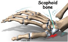
The scaphoid bone is one of the eight small bones that make up the “carpal bones” of the wrist. There are two rows of bones, one closer to the forearm (proximal row) and the other closer to
 the hand (distal row). The scaphoid bone is unique in that it links the two rows together (see Figure 1). This puts it at extra risk for injury, which accounts for it being the most commonly fractured carpal bone.
How do scaphoid fractures occur?
Fractures of the scaphoid occur most commonly from a fall on the outstretched hand.
 Usually it hurts at first, but the pain may improve quickly, over the course of days or weeks.
 Bruising is rare, and there is usually no visible deformity and only minimal swelling. 
Since there is no deformity, many people with this injury mistakenly assume that
 they have just sprained their wrist, leading to a delay in seeking evaluation. 
It is common for people who have fractured this bone to not become aware
 of it until months or years after the event.
Diagnosis of scaphoid fractures
Scaphoid fractures are most commonly diagnosed by x-rays of the wrist. However, 
when the fracture is not displaced, x-rays taken early (first week) may appear negative.
  A non-displaced scaphoid fracture could thus be incorrectly diagnosed as a “sprain.” 
Therefore a patient who has significant tenderness directly over the scaphoid bone 
(which is located in the hollow at the thumb side of the wrist, or “snuffbox”) 
should be suspected of having a scaphoid fracture and be splinted (see Figure 2). 
 An X-ray a couple of weeks later may then more clearly reveal the fracture. 
 In questionable cases, MRI scan, CT scan, or bone scan may be used to help
 diagnose an acute scaphoid fracture.  CT scan and/or MRI are also used to
 assess fracture displacement and configuration. Until a definitive diagnosis
 is made, the patient should remain splinted to prevent movement of a
 possible fracture.
Treatment of scaphoid fractures
If the fracture is non-displaced, it can be treated by immobilization in a cast
 that usually covers the forearm, hand, and thumb, and sometimes includes
 the elbow for the first phase of immobilization. Healing time in a cast can
 range from 6- 10 weeks and even longer. This is because the blood supply
 to the bone is variable and can be disrupted by the fracture, impairing bony
 healing. Part of the bone might even die after fracture due to loss of its 
blood supply, particularly in the proximal third of the bone, the part closest
 to the forearm. If the fracture is in this zone, or if it is at all displaced, 
surgery is more likely to be recommended. With surgery, a screw or pins
 are inserted to stabilize the fracture, sometimes with a bone graft to help
 heal the bone (see Figure 3a,3b).  Surgery to place a screw may also be
 recommended in non-displaced cases to avoid prolonged casting.
Complications of scaphoid fractures
Non-union: If a scaphoid fracture goes unrecognized, it often will not heal. 
Sometimes, even with treatment, it may not heal because of poor blood 
supply(seeFigure 3c). Over time, the abnormal motion and collapse of the
 bone fragments may lead to mal-alignment within the wrist and subsequent
 arthritis. If caught before arthritis has developed, surgery may be performed to
 try to get the scaphoid to heal.


Avascular necrosis:  A portion of the scaphoid may die because of lack of blood supply, leading to collapse of the bone and later arthritis.  Fractures in the proximal one third of the bone, the part closest to the forearm, are more vulnerable to this complication.  Again, if arthritis has not developed, surgery to try to stabilize the fracture and restore circulation to the bone may be attempted. (see.Figure 4)

Post-traumatic arthritis: If arthritis has already developed, salvage-type procedures may be considered, such as removal of degenerated bone or partial or complete fusion of the wrist joint.


Figure 1:  The scaphoid bone is unique in that it spans the two rows of wrist bones making it more prone to injury from a fall in full extension


Figure 2:  Significant tenderness directly over the scaphoid bone (which is located in the hollow at the thumb side of the wrist).


Figure 3a,3b:  A screw or pins are placed to stabilize the fracture.

Figure 3c.  The scaphoid has a varialbe pattern of blood supply that may predispose it to AVN and delayed healing

Figure 4. An MRI may be used to diagnose Avascular Necrosis (AVN) after a scaphoid fracture has not healed


parts © 2011 American Society for Surgery of the Hand. Developed by the ASSH Public Education Committee and Modified and supplemented for this blog by www.handcttr.com

Saturday, August 27, 2011

New Procedures Revolutionize Treatment of Dupuytren’s Disease


Helping Hands New Procedures Revolutionize Treatment of Dupuytren’s Disease BY DAN CHASE

July 2011

From left, Drs. Jeffrey Wint, Bruce Wintman, and Richard Martin at the Hand Center of Western Massachusetts.
Helping Hands
New Procedures Revolutionize Treatment of Dupuytren’s Disease
 BY DAN CHASE

It has been called many names, from the ‘Viking Disease,’ due to its prevalence among people of Northern European ancestry, to palmar fibromatosis. But to most patients and doctors, it is named for the French surgeon Baron Guillaume Dupuytren, who described an early treatment for the condition.
Dupuytren’s disease is a contraction of the fingers due to abnormal thickening of tissue. At the Hand Center of Western Massachusetts in Springfield, Drs. Richard Martin, Jeffrey Wint, and Bruce Wintman told HCN that the treatment of the disease has just been given a big helping hand. Two of them, in fact, in the form of new procedures that do nothing short of transform the way patients are given relief.
To get a better understanding of the mysteries of Dupuytren’s, which doesn’t have any specifically known causes — only a series of predispositions in patients — Wintman described the symptoms.
“There are different types of tissue in the hand,” he explained. “There’s skin, muscle, bone, and also a tissue that strands throughout the soft parts to give it form, called the fascia. In Dupuytren’s, your body makes a little bit too much of it, and an abnormal type of it at that.
“As a result,” he continued, “you start to get nodules in the palm into the fingers, and sometimes these can coalesce into a cord of tissue that can actually curl the finger down. That cord can be so thick that you can’t actually straighten it out on your own, even though the joints are normal and tendons are working. That abnormal fascial cord is flexing the fingers down. And that’s what patients complain about, that inability to straighten out those fingers.”
There is no pain particular to the abnormal fascia, but the lack of mobility can lead to problems with work and quality of life.
“It can become so cumbersome, and for some people can become so severe,” Wint added, “that we’ve had some patients say, ‘if you can’t do anything for it, cut the hand off. I just can’t do anything with it.’ That’s the extreme, though. Here, we tend to see anything from simple nodules to cords.”
Historically, the treatment for Dupuytren’s involved an open surgical procedure to remove the abnormal tissue, a process which also involves several months of physical therapy to restore mobility in the hand. However, new developments in the treatment of this condition have severed that downtime to the extent that a patient can have one of two new procedures — done in the office and not the surgical theater — and have immediate results with little aftereffect.
“In some cases,” Wintman said, “a patient can have one of these procedures during the week and be golfing by the weekend.”
This month, HCN sat down with the doctors of the Hand Center to find out what these new processes are, and how they are giving an important helping hand to the treatment of a life-altering condition.
General Contractor
Dupuytren’s is, as mentioned, a bit of a mystery, for there are no proven causes. But doctors say there are certain risk factors, including geographic ethnicity, age (over 40), a history of alcohol use, and, perhaps most importantly, those with a family history of the condition. While the disease is most commonly seen in men, it is not uncommon for women to contract it.
When the first signs of Dupuytren’s are noticed, the tendency is not to take immediate action, Martin said.
“If you get a little nodule, a little bump, there’s not a lot that needs to be done,” he explained. “However, when that becomes a cord, that’s the time when most people feel the need to do something about it. In years past, traditionally we would wait on treatment until it became a significant problem for the individual.”
Wintman said the “flat-table test” is a point of entry for his patients. “It’s usually not a pain situation whereby someone will want to seek treatment, but rather a lifestyle condition,” he explained. “If you can’t lay your hand flat on the table, then you may wish to proceed with treatment.”
For many years, what happened next was a pretty straightforward surgical process. If the nodule or cord was not contracting the fingers, nothing would be done, but the affected areas were kept under observation to monitor any marked development. If the progress contracted the fingers significantly, the hand would be incised, and the abnormal tissue cord removed. Wint said that this was a straightforward surgical procedure.
“The problem isn’t taking that cord out,” he explained. “That’s fairly easy to do. After an hour of surgery you can remove it. The problem is what happens to the hand afterwards. If it’s used to being bent, then the skin may be inefficient — it’s used to being pulled down. The tendons may not be springy right away, and the joints may want to reassume that bent position. So, someone who has a simple surgical removal to get rid of that cord needs to now go into a splint, sometimes full-time, up to a month, and into rehabilitation for three to six months after.”
Recurrence rates are a fact of life for patients with Dupuytren’s, and even with full removal of the contracting cord, the abnormal tissue can return in several years’ time. Also, if the original surgery involved the removal of a nodule, there’s a risk of not excising enough tissue, which Wint said could cause the abnormal tissue to return with increased vigor.
Handle with Care
While open surgery had been the only means for patients suffering this condition to return functionality to their fingers and hands, science has come to their rescue.
Two new procedures, as differentiated from surgery since they can be done in physicians’ offices, have come to the market within the last five years. One of these new techniques, a collagenase injection, was made available only one year ago.
That drug is called Xiaflex, and Wintman explained the process.
“You give a small shot of this enzyme in one area in the cord, and it eats through the collagen which the Dupuytren’s fascia is made from,” he said. “So it dissolves through the collagen, and then disrupts the cord at that one level. You come back the next day for manipulation, and the finger can go straight at that level after the manipulation.”
The second process is called needle aponeurotomy (NA), and essentially is a micro-perforation of the cord.
“We take a needle and actually make a series of puncture wounds along the length of the cord that’s keeping the finger bent down,” Martin said. “This breaks up the cord at various levels using the needle, almost like a tiny scalpel. The cord then actually breaks apart at these different levels. And then, the finger can straighten out. This doesn’t remove the tissue; you’re just breaking it up.
“As a result,” he continued, “afterward there are just a series of exterior puncture wounds that need to heal, typically over the course of five to seven days. Patients go home with some Band-Aids and an ice pack, and they’re moving and using their hand right away. Typically they don’t need a splint, although we might give them one to reinforce the gains we’ve made. But that is rare.”
Choosing between the new procedures comes down, ultimately, to dollars and sense: often an insurance carrier might cover one but not the other.
Also, the NA can involve several affected areas in the hand on one visit, whereas the Xiaflex must be administered only in one area every 30 days.
“So if someone comes in with three fingers affected, that’s a bit of a stretch,” Wintman said. “But some people want to try the medication over the four months.”
However, he added, “a patient might have co-morbidities. They might be on a blood thinner, and you wouldn’t want to give them an injection of an enzyme that eats tissue, because there would be too much swelling. Some people may have skin issues and not want to be exposed.”
But it can come down to a patient’s word-of-mouth referral to determine one over the other, Wint said.
Around the Bend
Sadly, science has not cured Dupuytren’s once and for all. The same recurrence rates for the abnormal tissues does still apply to these two new procedures, and, in fact, the time until reappearance of nodules and cords is often just a few years’ time. But, doctors agree, the ease with which both Xiaflex and NA can be administered should allay any fears of a return visit.
But has science sounded the death knell for the surgical open procedure?
“Well, it does reduce that frequency significantly,” Wint said. “Since that used to be the only solution, it was that or nothing. I had some people who, because of their work, really couldn’t take three to four months off. And you had to try to prepare people, also, because it could have even been longer.”
Martin said there are patients who still say, “I want it out,” and that does mean surgery. And for a patient with a history of severe Dupuytren’s, or situations involving prior complex surgeries, the new procedures might not offer any assistance.
“Their anatomy might be markedly distorted; they may have had skin grafts done elsewhere,” Wintman said. “Also, you can have contractures of the joints — the joint itself is scarred in. It’s not that you can feel a clearly defined cord anymore, but there might be scar tissue that keeps the fingers contracted. And, unfortunately, you really can’t do anything with that in the new procedures.”
But one thing the two new techniques will hopefully bring about is a future whereby doctors don’t see such severe cases of Dupuytren’s. “As more and more people read about these procedures, or hear about them from friends, they’ll realize that the need for the surgery will be less and less,” Martin said.
His colleagues nodded in agreement when Wintman said, “it’s incredibly satisfying for us, because when the patient leaves the office, they will have noticeable results. They walk in with a deficit, and they leave here with straight fingers.”

Wednesday, August 17, 2011

TENNIS ELBOW or LATERAL EPICONDYLITIS

Tennis Elbow: treatment (Lateral Epicondylitis)treatment

Bookmark and Share
taken and modified from the American Academy of Orthopedic Surgery (AAOS) website
for additional information taken from ASSH website  to see our office website click here
DescriptionTennis elbow is a degenerative condition of the tendon fibers that attach on the bony prominence (epicondyle) on the outside (lateral side) of the elbow. The tendons involved are responsible for anchoring the muscles that extend or lift the wrist and hand (see Figure 1).


Risk Factors/Prevention
Tennis elbow happens mostly in patients between the ages of 30 years to 50 years. It can occur in any age group. Tennis elbow can affect as many as half of athletes in racquet sports. However, most patients with tennis elbow are not active in racquet sports. Most of the time, there is not a specific traumatic injury before symptoms start. Many individuals with tennis elbow are involved in work or recreational activities that require repetitive and vigorous use of the forearm muscles (see Table 1). Some patients develop tennis elbow without any specific recognizable activity leading to symptoms.


Symptoms
Patients often complain of severe, burning pain on the outside part of the elbow. In most cases, the pain starts in a mild and slow fashion. It gradually worsens over weeks or months. The pain can be made worse by pressing on the outside part of the elbow or by gripping or lifting objects. Lifting even very light objects (such as a small book or a cup of coffee) can lead to significant discomfort. In more severe cases, pain can occur with simple motion of the elbow joint. Pain can radiate to the forearm.
To diagnose tennis elbow, tell the doctor your complete medical history. He or she will perform a physical examination.
  • The doctor may press directly on the bony prominence on the outside part of the elbow to see if it causes pain.
  •  
  • The doctor may also ask you to lift the wrist or fingers against pressure to see if that causes pain.
X-rays may be necessary.  Xrays can reveal the presence of calcification or arthritis that may also affect the elbow joint. Rarely, MRI (magnetic resonance imaging) scans may be used to show changes in the tendon at the site of attachment onto the bone for acute high energy injuries

Treatment Options
In many cases  when there are fairly severe symptoms a simultaneous-three pronged approach,  (1) use of anti-inflammatories,  (2) orthotic bracing and  (3)injection  at the same time work more efficaciously and can save time in treatment and offers a more rapid resolution of symptoms. Sometimes, when conditions are right a more slow sequential treatment is offered. This takes longer but may be more suitable for those with minimal symptoms
In most cases, nonoperative treatment should be tried before surgery. Pain relief is the main goal in the first phase of treatment. The doctor may tell you to stop any activities that cause symptoms. You may need to apply ice to the outside part of the elbow. You may need to take acetaminophen or an anti-inflammatory medication for pain relief.
Orthotics can help diminish symptoms of tennis elbow. The doctor may want you to use counterforce braces and wrist splints. These can reduce symptoms by resting the muscles and tendons (see Figure 2).


Symptoms should improve significantly within four weeks to six weeks. If not, the next step may be a corticosteroid injection around the outside of the elbow. This can be very helpful in reducing pain. Corticosteroids are relatively safe medications. Most of their side effects (i.e., further degeneration of the tendon and wasting of the fatty tissue below the skin) occur after multiple injections. Avoid repeated injections (more than two or three in a specific site).As noted above some patients symptoms are severe enough to warrant a simulateous-three pronged approach at the time of initial consultation.

After pain is relieved, the next phase of treatment starts. Modifying activities can help make sure that symptoms do not come back. The doctor may want you to do physical therapy. This may include stretching and range of motion exercises and gradual strengthening of the affected muscles and tendons (see Figure 3). Physical therapy can help complete recovery and give you back a painless and normally functioning elbow.  In many cases however PT is not needed once pain has subsided.  Nonoperative treatment is successful in approximately 85 percent to 90 percent of patients with tennis elbow.
Treatment Options: Surgical
Surgery is considered only in patients who have incapacitating* pain that does not get better after  non-operative treatment.
*The difficult question to answer is:  What is incapacitating pain?  Very often what affects one person may not be significant for another.  This is especially true for those engaged in athletics or jobs that require very specific motions or activities.  Thus the indications for surgery are relative in most and absolute in few.

The surgical procedure involves removing diseased tendon tissue and reattaching normal tendon tissue to bone (see Figure 4). The procedure is an outpatient surgery; you do not need to stay in the hospital overnight. It can be performed under regional or general anesthesia.


Technique for surgical treatment of lateral epicondylitis. A, Skin incision over the lateral epicondyle. B, Distal reflection of the extensor mechanism exposing the lateral compartment of the elbow. C, Excision of pathologic tissue from the underside of the extensor mechanism. D, Decortication of the lateral epicondyle. E, Drilling of two V-shaped tunnels within the lateral epicondyle. F, Reattachment of the extensor mechanism to the lateral epicondyle. G, Side-to-side repair of the extensor tendon mechanism. 
 *Note: steps E and F are not often needed except in cases where large *tears exist.

  • Most commonly, the surgery is performed through a small incision over the bony prominence on the outside of the elbow.

So far, no significant benefits have been found to using an arthroscopic method over the  traditional open incision.
After surgery, the elbow is placed in a small brace and the patient is sent home. About one week later, the sutures and splint are removed. Then exercises are started to stretch the elbow and restore range of motion. Light, gradual strengthening exercises are started two months after surgery. The doctor will tell you when you can return to athletic activity. This is usually approximately four months to six months after surgery. Tennis elbow surgery is considered successful in approximately 90 percent of patients.
Persistent pain can be from associated arthritis in the elbow, nerve compression, muscular fatigue or residual bone pain.  While most patients return to full activity after tennis elbow surgery, many still relate that there is aching pain that persists at times.  Overall, however, most are satisfied and have a significant improvement and much less pain than prior to surgery

November 2004

Author Information (Please note that although the original document has been modified  we wish to grant the original author from the AAOS credit.)

 Hit Counter

Friday, August 12, 2011

Carpal Boss of the WRIST and HAND


Click to visit our website
Carpal Boss
  Bookmark and Share
Clinical Characteristics
The dorsal wrist ganglion is most often confused with the carpal boss, so named by the French physician Foille.  The carpal boss is an osteoarthritic spur that develops at the base of the second and/or third carpometacarpal joints. (figure 1) A firm, bony, nonmobile, tender mass is visible and palpable at the base of the carpometacarpal joints, especially when the wrist is volar flexed.
Radiologically, the mass is best visualized with the hand in 30 to 40 degrees supination and  
 20 to 30 degrees ulnar deviation ("carpal boss view")( figure 3).


figure1


The boss is more common in women (2:1), in the right hand (2:1), and between the third and fourth decades.  The mass may be asymptomatic, but the patient may complain of considerable pain and aching.  A small ganglion is associated with  the carpal boss in 30 percent of cases, adding to its confusion with the more common dorsal wrist ganglion. 
Injection to the ganglion or to the cmc joint may  be used to reduce pain and irritation.  This may be combined with splinting and anti inflammatory medication and avoidance of trauma to the back of the hand\
If symptoms persist at times surgery may be offered. (figure 2) Surgery may involve the removal of the prominent bone, the excision of an associated ganglion or cyst and at times involves tenosynovectomy or tenolysis of adjacent affected tendons.  What occurs during surgery may depend upon the preoperative findings as well as the surgical intra operative findings
figure 2
The most common complication is the persistence of a mass because of excision of the ganglion alone or inadequate excision of the osteophytes.  Pain will persist unless all abnormal abutting surfaces have been excised.  Dorsal wrist ganglions can present over the carpometacarpal joints and must be distinguished from the carpal boss with its own associated ganglion.  Avoidance of injury to branches of the radial and ulnar sensory nerves is again stressed.            
figure 3

Wednesday, July 20, 2011

Helping Hands New Procedures Revolutionize Treatment of Dupuytren’s Disease BY DAN CHASE

July 2011

From left, Drs. Jeffrey Wint, Bruce Wintman, and Richard Martin at the Hand Center of Western Massachusetts.
Helping Hands
New Procedures Revolutionize Treatment of Dupuytren’s Disease
 BY DAN CHASE

It has been called many names, from the ‘Viking Disease,’ due to its prevalence among people of Northern European ancestry, to palmar fibromatosis. But to most patients and doctors, it is named for the French surgeon Baron Guillaume Dupuytren, who described an early treatment for the condition.
Dupuytren’s disease is a contraction of the fingers due to abnormal thickening of tissue. At the Hand Center of Western Massachusetts in Springfield, Drs. Richard Martin, Jeffrey Wint, and Bruce Wintman told HCN that the treatment of the disease has just been given a big helping hand. Two of them, in fact, in the form of new procedures that do nothing short of transform the way patients are given relief.
To get a better understanding of the mysteries of Dupuytren’s, which doesn’t have any specifically known causes — only a series of predispositions in patients — Wintman described the symptoms.
“There are different types of tissue in the hand,” he explained. “There’s skin, muscle, bone, and also a tissue that strands throughout the soft parts to give it form, called the fascia. In Dupuytren’s, your body makes a little bit too much of it, and an abnormal type of it at that.
“As a result,” he continued, “you start to get nodules in the palm into the fingers, and sometimes these can coalesce into a cord of tissue that can actually curl the finger down. That cord can be so thick that you can’t actually straighten it out on your own, even though the joints are normal and tendons are working. That abnormal fascial cord is flexing the fingers down. And that’s what patients complain about, that inability to straighten out those fingers.”
There is no pain particular to the abnormal fascia, but the lack of mobility can lead to problems with work and quality of life.
“It can become so cumbersome, and for some people can become so severe,” Wint added, “that we’ve had some patients say, ‘if you can’t do anything for it, cut the hand off. I just can’t do anything with it.’ That’s the extreme, though. Here, we tend to see anything from simple nodules to cords.”
Historically, the treatment for Dupuytren’s involved an open surgical procedure to remove the abnormal tissue, a process which also involves several months of physical therapy to restore mobility in the hand. However, new developments in the treatment of this condition have severed that downtime to the extent that a patient can have one of two new procedures — done in the office and not the surgical theater — and have immediate results with little aftereffect.
“In some cases,” Wintman said, “a patient can have one of these procedures during the week and be golfing by the weekend.”
This month, HCN sat down with the doctors of the Hand Center to find out what these new processes are, and how they are giving an important helping hand to the treatment of a life-altering condition.
General Contractor
Dupuytren’s is, as mentioned, a bit of a mystery, for there are no proven causes. But doctors say there are certain risk factors, including geographic ethnicity, age (over 40), a history of alcohol use, and, perhaps most importantly, those with a family history of the condition. While the disease is most commonly seen in men, it is not uncommon for women to contract it.
When the first signs of Dupuytren’s are noticed, the tendency is not to take immediate action, Martin said.
“If you get a little nodule, a little bump, there’s not a lot that needs to be done,” he explained. “However, when that becomes a cord, that’s the time when most people feel the need to do something about it. In years past, traditionally we would wait on treatment until it became a significant problem for the individual.”
Wintman said the “flat-table test” is a point of entry for his patients. “It’s usually not a pain situation whereby someone will want to seek treatment, but rather a lifestyle condition,” he explained. “If you can’t lay your hand flat on the table, then you may wish to proceed with treatment.”
For many years, what happened next was a pretty straightforward surgical process. If the nodule or cord was not contracting the fingers, nothing would be done, but the affected areas were kept under observation to monitor any marked development. If the progress contracted the fingers significantly, the hand would be incised, and the abnormal tissue cord removed. Wint said that this was a straightforward surgical procedure.
“The problem isn’t taking that cord out,” he explained. “That’s fairly easy to do. After an hour of surgery you can remove it. The problem is what happens to the hand afterwards. If it’s used to being bent, then the skin may be inefficient — it’s used to being pulled down. The tendons may not be springy right away, and the joints may want to reassume that bent position. So, someone who has a simple surgical removal to get rid of that cord needs to now go into a splint, sometimes full-time, up to a month, and into rehabilitation for three to six months after.”
Recurrence rates are a fact of life for patients with Dupuytren’s, and even with full removal of the contracting cord, the abnormal tissue can return in several years’ time. Also, if the original surgery involved the removal of a nodule, there’s a risk of not excising enough tissue, which Wint said could cause the abnormal tissue to return with increased vigor.
Handle with Care
While open surgery had been the only means for patients suffering this condition to return functionality to their fingers and hands, science has come to their rescue.
Two new procedures, as differentiated from surgery since they can be done in physicians’ offices, have come to the market within the last five years. One of these new techniques, a collagenase injection, was made available only one year ago.
That drug is called Xiaflex, and Wintman explained the process.
“You give a small shot of this enzyme in one area in the cord, and it eats through the collagen which the Dupuytren’s fascia is made from,” he said. “So it dissolves through the collagen, and then disrupts the cord at that one level. You come back the next day for manipulation, and the finger can go straight at that level after the manipulation.”
The second process is called needle aponeurotomy (NA), and essentially is a micro-perforation of the cord.
“We take a needle and actually make a series of puncture wounds along the length of the cord that’s keeping the finger bent down,” Martin said. “This breaks up the cord at various levels using the needle, almost like a tiny scalpel. The cord then actually breaks apart at these different levels. And then, the finger can straighten out. This doesn’t remove the tissue; you’re just breaking it up.
“As a result,” he continued, “afterward there are just a series of exterior puncture wounds that need to heal, typically over the course of five to seven days. Patients go home with some Band-Aids and an ice pack, and they’re moving and using their hand right away. Typically they don’t need a splint, although we might give them one to reinforce the gains we’ve made. But that is rare.”
Choosing between the new procedures comes down, ultimately, to dollars and sense: often an insurance carrier might cover one but not the other.
Also, the NA can involve several affected areas in the hand on one visit, whereas the Xiaflex must be administered only in one area every 30 days.
“So if someone comes in with three fingers affected, that’s a bit of a stretch,” Wintman said. “But some people want to try the medication over the four months.”
However, he added, “a patient might have co-morbidities. They might be on a blood thinner, and you wouldn’t want to give them an injection of an enzyme that eats tissue, because there would be too much swelling. Some people may have skin issues and not want to be exposed.”
But it can come down to a patient’s word-of-mouth referral to determine one over the other, Wint said.
Around the Bend
Sadly, science has not cured Dupuytren’s once and for all. The same recurrence rates for the abnormal tissues does still apply to these two new procedures, and, in fact, the time until reappearance of nodules and cords is often just a few years’ time. But, doctors agree, the ease with which both Xiaflex and NA can be administered should allay any fears of a return visit.
But has science sounded the death knell for the surgical open procedure?
“Well, it does reduce that frequency significantly,” Wint said. “Since that used to be the only solution, it was that or nothing. I had some people who, because of their work, really couldn’t take three to four months off. And you had to try to prepare people, also, because it could have even been longer.”
Martin said there are patients who still say, “I want it out,” and that does mean surgery. And for a patient with a history of severe Dupuytren’s, or situations involving prior complex surgeries, the new procedures might not offer any assistance.
“Their anatomy might be markedly distorted; they may have had skin grafts done elsewhere,” Wintman said. “Also, you can have contractures of the joints — the joint itself is scarred in. It’s not that you can feel a clearly defined cord anymore, but there might be scar tissue that keeps the fingers contracted. And, unfortunately, you really can’t do anything with that in the new procedures.”
But one thing the two new techniques will hopefully bring about is a future whereby doctors don’t see such severe cases of Dupuytren’s. “As more and more people read about these procedures, or hear about them from friends, they’ll realize that the need for the surgery will be less and less,” Martin said.
His colleagues nodded in agreement when Wintman said, “it’s incredibly satisfying for us, because when the patient leaves the office, they will have noticeable results. They walk in with a deficit, and they leave here with straight fingers.”