CHAT

Tuesday, November 29, 2011

SNOW BLOWER SAFETY TIP... (O. C. D. STICK)

BE PREPARED THIS WINTER PRACTICE SNOW BLOWER SAFETY

Snow Blower Safety

Conditions that are associated with a higher incidence of injuries, heavy wet snow exceeding 6 inches of accumulation and temperatures above 28 degrees Fahrenheit offer good opportunities to provide warning for the public. We need your help to reduce the incidence of these preventable injuries.

Recommendations for safe use of a jammed snow blower snow blowers include: (OCD STICK)

1. If the snow blower jams, immediately turn it OFF
2. Disengage the CLUTCH
3. DELAY...Wait 10 seconds after shutting of to allow Impeller Blades to stop rotating
4. Always use a STICK or broom handle to clear impacted snow. The stick most be strong enough to avoid breakage or eye injures can result from flying fragments.
5. Never put your hand near chute or around blades
6. Keep all shields in place. Do not remove safety devices on machine
7. Keep hands and feet away from moving parts
8. Keep a clear head, concentrate and ...
    Do not drink alcoholic beverages before using a snow blower

As physicians dedicated to the care of the Hand and Upper extremity we want to inform the public concerning the perils and pitfalls of improper snow blower use.  Physicians, nurses, allied health professionals and therapists who deal with these injuries live in fear of the first heavy wet snow of the season. Invariably injuries are seen despite general knowledge that these injuries occur. These safety tips cannot guarantee against injury but hopefully if you are reading these or even better spreading these, it is one more step towards preventing these types of injuries.

News organizations and weather services can help.

Conditions that are associated with a higher incidence of injuries, hay wet snow exceeding 6 inches of accumulation and temperatures above 28 degrees Fahrenheit offer good opportunities to provide warning for the public. We need your help to reduce the incidence of these preventable injuries.


Helping Hands New Procedures Revolutionize Treatment of Dupuytren’s Disease


 BY DAN CHASE

July 2011

From left, Drs. Jeffrey Wint, Bruce Wintman, and Richard Martin at the Hand Center of Western Massachusetts.
Helping Hands
New Procedures Revolutionize Treatment of Dupuytren’s Disease
 BY DAN CHASE

It has been called many names, from the ‘Viking Disease,’ due to its prevalence among people of Northern European ancestry, to palmar fibromatosis. But to most patients and doctors, it is named for the French surgeon Baron Guillaume Dupuytren, who described an early treatment for the condition.
Dupuytren’s disease is a contraction of the fingers due to abnormal thickening of tissue. At the Hand Center of Western Massachusetts in Springfield, Drs. Richard Martin, Jeffrey Wint, and Bruce Wintman told HCN that the treatment of the disease has just been given a big helping hand. Two of them, in fact, in the form of new procedures that do nothing short of transform the way patients are given relief.
To get a better understanding of the mysteries of Dupuytren’s, which doesn’t have any specifically known causes — only a series of predispositions in patients — Wintman described the symptoms.
“There are different types of tissue in the hand,” he explained. “There’s skin, muscle, bone, and also a tissue that strands throughout the soft parts to give it form, called the fascia. In Dupuytren’s, your body makes a little bit too much of it, and an abnormal type of it at that.
“As a result,” he continued, “you start to get nodules in the palm into the fingers, and sometimes these can coalesce into a cord of tissue that can actually curl the finger down. That cord can be so thick that you can’t actually straighten it out on your own, even though the joints are normal and tendons are working. That abnormal fascial cord is flexing the fingers down. And that’s what patients complain about, that inability to straighten out those fingers.”
There is no pain particular to the abnormal fascia, but the lack of mobility can lead to problems with work and quality of life.
“It can become so cumbersome, and for some people can become so severe,” Wint added, “that we’ve had some patients say, ‘if you can’t do anything for it, cut the hand off. I just can’t do anything with it.’ That’s the extreme, though. Here, we tend to see anything from simple nodules to cords.”
Historically, the treatment for Dupuytren’s involved an open surgical procedure to remove the abnormal tissue, a process which also involves several months of physical therapy to restore mobility in the hand. However, new developments in the treatment of this condition have severed that downtime to the extent that a patient can have one of two new procedures — done in the office and not the surgical theater — and have immediate results with little aftereffect.
“In some cases,” Wintman said, “a patient can have one of these procedures during the week and be golfing by the weekend.”
This month, HCN sat down with the doctors of the Hand Center to find out what these new processes are, and how they are giving an important helping hand to the treatment of a life-altering condition.
General Contractor
Dupuytren’s is, as mentioned, a bit of a mystery, for there are no proven causes. But doctors say there are certain risk factors, including geographic ethnicity, age (over 40), a history of alcohol use, and, perhaps most importantly, those with a family history of the condition. While the disease is most commonly seen in men, it is not uncommon for women to contract it.
When the first signs of Dupuytren’s are noticed, the tendency is not to take immediate action, Martin said.
“If you get a little nodule, a little bump, there’s not a lot that needs to be done,” he explained. “However, when that becomes a cord, that’s the time when most people feel the need to do something about it. In years past, traditionally we would wait on treatment until it became a significant problem for the individual.”
Wintman said the “flat-table test” is a point of entry for his patients. “It’s usually not a pain situation whereby someone will want to seek treatment, but rather a lifestyle condition,” he explained. “If you can’t lay your hand flat on the table, then you may wish to proceed with treatment.”
For many years, what happened next was a pretty straightforward surgical process. If the nodule or cord was not contracting the fingers, nothing would be done, but the affected areas were kept under observation to monitor any marked development. If the progress contracted the fingers significantly, the hand would be incised, and the abnormal tissue cord removed. Wint said that this was a straightforward surgical procedure.
“The problem isn’t taking that cord out,” he explained. “That’s fairly easy to do. After an hour of surgery you can remove it. The problem is what happens to the hand afterwards. If it’s used to being bent, then the skin may be inefficient — it’s used to being pulled down. The tendons may not be springy right away, and the joints may want to reassume that bent position. So, someone who has a simple surgical removal to get rid of that cord needs to now go into a splint, sometimes full-time, up to a month, and into rehabilitation for three to six months after.”
Recurrence rates are a fact of life for patients with Dupuytren’s, and even with full removal of the contracting cord, the abnormal tissue can return in several years’ time. Also, if the original surgery involved the removal of a nodule, there’s a risk of not excising enough tissue, which Wint said could cause the abnormal tissue to return with increased vigor.
Handle with Care
While open surgery had been the only means for patients suffering this condition to return functionality to their fingers and hands, science has come to their rescue.
Two new procedures, as differentiated from surgery since they can be done in physicians’ offices, have come to the market within the last five years. One of these new techniques, a collagenase injection, was made available only one year ago.
That drug is called Xiaflex, and Wintman explained the process.
“You give a small shot of this enzyme in one area in the cord, and it eats through the collagen which the Dupuytren’s fascia is made from,” he said. “So it dissolves through the collagen, and then disrupts the cord at that one level. You come back the next day for manipulation, and the finger can go straight at that level after the manipulation.”
The second process is called needle aponeurotomy (NA), and essentially is a micro-perforation of the cord.
“We take a needle and actually make a series of puncture wounds along the length of the cord that’s keeping the finger bent down,” Martin said. “This breaks up the cord at various levels using the needle, almost like a tiny scalpel. The cord then actually breaks apart at these different levels. And then, the finger can straighten out. This doesn’t remove the tissue; you’re just breaking it up.
“As a result,” he continued, “afterward there are just a series of exterior puncture wounds that need to heal, typically over the course of five to seven days. Patients go home with some Band-Aids and an ice pack, and they’re moving and using their hand right away. Typically they don’t need a splint, although we might give them one to reinforce the gains we’ve made. But that is rare.”
Choosing between the new procedures comes down, ultimately, to dollars and sense: often an insurance carrier might cover one but not the other.
Also, the NA can involve several affected areas in the hand on one visit, whereas the Xiaflex must be administered only in one area every 30 days.
“So if someone comes in with three fingers affected, that’s a bit of a stretch,” Wintman said. “But some people want to try the medication over the four months.”
However, he added, “a patient might have co-morbidities. They might be on a blood thinner, and you wouldn’t want to give them an injection of an enzyme that eats tissue, because there would be too much swelling. Some people may have skin issues and not want to be exposed.”
But it can come down to a patient’s word-of-mouth referral to determine one over the other, Wint said.
Around the Bend
Sadly, science has not cured Dupuytren’s once and for all. The same recurrence rates for the abnormal tissues does still apply to these two new procedures, and, in fact, the time until reappearance of nodules and cords is often just a few years’ time. But, doctors agree, the ease with which both Xiaflex and NA can be administered should allay any fears of a return visit.
But has science sounded the death knell for the surgical open procedure?
“Well, it does reduce that frequency significantly,” Wint said. “Since that used to be the only solution, it was that or nothing. I had some people who, because of their work, really couldn’t take three to four months off. And you had to try to prepare people, also, because it could have even been longer.”
Martin said there are patients who still say, “I want it out,” and that does mean surgery. And for a patient with a history of severe Dupuytren’s, or situations involving prior complex surgeries, the new procedures might not offer any assistance.
“Their anatomy might be markedly distorted; they may have had skin grafts done elsewhere,” Wintman said. “Also, you can have contractures of the joints — the joint itself is scarred in. It’s not that you can feel a clearly defined cord anymore, but there might be scar tissue that keeps the fingers contracted. And, unfortunately, you really can’t do anything with that in the new procedures.”
But one thing the two new techniques will hopefully bring about is a future whereby doctors don’t see such severe cases of Dupuytren’s. “As more and more people read about these procedures, or hear about them from friends, they’ll realize that the need for the surgery will be less and less,” Martin said.
His colleagues nodded in agreement when Wintman said, “it’s incredibly satisfying for us, because when the patient leaves the office, they will have noticeable results. They walk in with a deficit, and they leave here with straight fingers.”

SCAPHOID FRACTURES

SCAPHOID FRACTURES of the WRIST

Scaphoid Fractures

What are scaphoid fractures?
The scaphoid bone is one of the eight small bones that make up the “carpal bones” of the wrist. There are two rows of bones, one closer to the forearm (proximal row) and the other closer to
 the hand (distal row). The scaphoid bone is unique in that it links the two rows together (see Figure 1). This puts it at extra risk for injury, which accounts for it being the most commonly fractured carpal bone.
How do scaphoid fractures occur?
Fractures of the scaphoid occur most commonly from a fall on the outstretched hand.
 Usually it hurts at first, but the pain may improve quickly, over the course of days or weeks.
 Bruising is rare, and there is usually no visible deformity and only minimal swelling. 
Since there is no deformity, many people with this injury mistakenly assume that
 they have just sprained their wrist, leading to a delay in seeking evaluation. 
It is common for people who have fractured this bone to not become aware
 of it until months or years after the event.
Diagnosis of scaphoid fractures
Scaphoid fractures are most commonly diagnosed by x-rays of the wrist. However, 
when the fracture is not displaced, x-rays taken early (first week) may appear negative.
  A non-displaced scaphoid fracture could thus be incorrectly diagnosed as a “sprain.” 
Therefore a patient who has significant tenderness directly over the scaphoid bone 
(which is located in the hollow at the thumb side of the wrist, or “snuffbox”) 
should be suspected of having a scaphoid fracture and be splinted (see Figure 2). 
 An X-ray a couple of weeks later may then more clearly reveal the fracture. 
 In questionable cases, MRI scan, CT scan, or bone scan may be used to help
 diagnose an acute scaphoid fracture.  CT scan and/or MRI are also used to
 assess fracture displacement and configuration. Until a definitive diagnosis
 is made, the patient should remain splinted to prevent movement of a
 possible fracture.
Treatment of scaphoid fractures
If the fracture is non-displaced, it can be treated by immobilization in a cast
 that usually covers the forearm, hand, and thumb, and sometimes includes
 the elbow for the first phase of immobilization. Healing time in a cast can
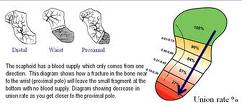
 range from 6- 10 weeks and even longer. This is because the blood supply
 to the bone is variable and can be disrupted by the fracture, impairing bony
 healing. Part of the bone might even die after fracture due to loss of its 
blood supply, particularly in the proximal third of the bone, the part closest
 to the forearm. If the fracture is in this zone, or if it is at all displaced, 
surgery is more likely to be recommended. With surgery, a screw or pins
 are inserted to stabilize the fracture, sometimes with a bone graft to help
 heal the bone (see Figure 3a,3b).  Surgery to place a screw may also be
 recommended in non-displaced cases to avoid prolonged casting.
Complications of scaphoid fractures
Non-union: If a scaphoid fracture goes unrecognized, it often will not heal. 
Sometimes, even with treatment, it may not heal because of poor blood 
supply(seeFigure 3c). Over time, the abnormal motion and collapse of the
 bone fragments may lead to mal-alignment within the wrist and subsequent
 arthritis. If caught before arthritis has developed, surgery may be performed to
 try to get the scaphoid to heal.


Avascular necrosis:  A portion of the scaphoid may die because of lack of blood supply, leading to collapse of the bone and later arthritis.  Fractures in the proximal one third of the bone, the part closest to the forearm, are more vulnerable to this complication.  Again, if arthritis has not developed, surgery to try to stabilize the fracture and restore circulation to the bone may be attempted. (see.Figure 4)

Post-traumatic arthritis: If arthritis has already developed, salvage-type procedures may be considered, such as removal of degenerated bone or partial or complete fusion of the wrist joint.


Figure 1:  The scaphoid bone is unique in that it spans the two rows of wrist bones making it more prone to injury from a fall in full extension


Figure 2:  Significant tenderness directly over the scaphoid bone (which is located in the hollow at the thumb side of the wrist).


Figure 3a,3b:  A screw or pins are placed to stabilize the fracture.

Figure 3c.  The scaphoid has a varialbe pattern of blood supply that may predispose it to AVN and delayed healing

Figure 4. An MRI may be used to diagnose Avascular Necrosis (AVN) after a scaphoid fracture has not healed


parts © 2011 American Society for Surgery of the Hand. Developed by the ASSH Public Education Committee and Modified and supplemented for this blog by www.handcttr.com


BE PREPARED THIS WINTER PRACTICE SNOW BLOWER SAFETY

Snow Blower Safety

Conditions that are associated with a higher incidence of injuries, hay wet snow exceeding 6 inches of accumulation and temperatures above 28 degrees Fahrenheit offer good opportunities to provide warning for the public. We need your help to reduce the incidence of these preventable injuries.

Recommendations for safe use of a jammed snow blower snow blowers include: (OCD STICK)

1. If the snow blower jams, immediately turn it OFF
2. Disengage the CLUTCH
3. DELAY...Wait 10 seconds after shutting of to allow Impeller Blades to stop rotating
4. Always use a STICK or broom handle to clear impacted snow. The stick most be strong enough to avoid breakage or eye injures can result from flying fragments.
5. Never put your hand near chute or around blades
6. Keep all shields in place. Do not remove safety devices on machine
7. Keep hands and feet away from moving parts
8. Keep a clear head, concentrate and ...
    Do not drink alcoholic beverages before using a snow blower

As physicians dedicated to the care of the Hand and Upper extremity we want to inform the public concerning the perils and pitfalls of improper snow blower use.  Physicians, nurses, allied health professionals and therapists who deal with these injuries live in fear of the first heavy wet snow of the season. Invariably injuries are seen despite general knowledge that these injuries occur. These safety tips cannot guarantee against injury but hopefully if you are reading these or even better spreading these, it is one more step towards preventing these types of injuries.

News organizations and weather services can help.

Conditions that are associated with a higher incidence of injuries, hay wet snow exceeding 6 inches of accumulation and temperatures above 28 degrees Fahrenheit offer good opportunities to provide warning for the public. We need your help to reduce the incidence of these preventable injuries.


MALLET FINGER (baseball finger)

MALLET FINGER (baseball finger)

MALLET FINGER (BASEBALL FINGER)


A mallet finger occurs when the extensor tendon at the tip of a finger ruptures. The rupture of this tendon can involve the tendon alone, be associated with a small bone fragment or fracture or can be associated with a fracture that requires significant care.

The force applied to the finger can come from something as simple as tucking in a bed sheet or can come from a direct blow to the end of a finger. Mallet finger has also been known as baseball finger.

A mallet finger often begins with pain at the distal joint of the finger.  At times there is an immediate loss of motion while at other times the finger seems to stay straight for a while and only later starts to lose its ability to be extended actively at the tip.  At times there is an injuries are typically closed in that the skin and nail is intact but at times there is an injury to the skin or nail bed as well. In severe cases the injury is associated with an open injury to the joint or bone, a so called open or compound fracture.

In adults the injury can involve the joint surface.  In children it can involves the growth plate or physis.

The diagnosis is often made based upon the type of injury and the appearance of the finger.  The fingertip will droop down and there is a loss of active motion.  Often the finger can be passively pushed up to straighten it but the independent active motion to extend the digit at the tip has been lost


Mallet finger.

X-rays are often taken to further delineate the injury and see how much if any bone, joint or growth plate is involved

Treatment depends largely upon the extent the soft tissue and underlying boney injury.

Tendon rupture without bone injury

Most of these mallet finger injuries can be treated with splinting.  The splint can be applied in a variety of ways depending upon the injury.  Typically the split is left in place full time for six to eight weeks with a time for part time splinting after that depending upon what daily activity is done by the patient with a typical part time period of 3 – 4 weeks.  In some situations pinning of the joint is used rather than a splint

Tendon rupture with a small bone fragment

These injuries typically are treated like non-boney injuries


Types of splints used to treat mallet finger. A, Dorsal aluminum splint. B, Commercial splint.
Reproduced with permission from Culver JE Jr: Office management of athletic injuries of the hand and wrist. Instr Course Lect 1989;38:473-482.


Tendon rupture with a large bone fragment involving the joint.


These injuries may respond to splinting and splinting is often used however a small bump may always be present a t the joint.  At times if the doctor feels that that the bone fragment is large enough and the joint may be unstable surgery may be offered.  During surgery pins or small screws may be used and the joint itself may be pinned to prevent motion during the healing process.

Above: X-rays showing fracture at the insertion of the extensor tendon. In the first image on the left the fragment is displaced.  This will heal with a bump but will be able to be treated with a splint.  IN the image on the right the joint has subluxed.  This will  need to have surgical repair.
In adults with severe open injury more immediate surgery may also be offered

Children

In children the doctor needs to differentiate between these injuries that require reduction or realignment of the bone without surgery and those who may have a portion of the nail bed significantly torn or retained within the fracture site or growth plate.  Often children will not have a tendon injury but a fracture through the physis which appears to be a mallet injury.  X-rays often will reveal this.


Mallet deformity from a fracture across the growth plate in a child is different than the adult fracture or tendon avulsion

Late or Delayed Treatment in adults

Delayed treatment of mallet finger deformity may consist of splinting initially and at times surgical methods are offered to correct chronic deformities and other associated joint and tendon problems that may accompany the chronic situation

Results

Most mallet fingers heal well, although often there is a slight loss of full extension. The slight extension loss typically has no effect on hand of finger function, but if left untreated it can cause other issues to occur in the finger due to tendon imbalance. While treatment of a closed mallet finger is not an acute emergency, the improper, partial or untreated injury can lead to further problems such as a swan neck deformity.

 Swan neck deformity from tendon imbalance and laxity at the proximal joint.

EXAMPLE: A  TYPICAL TREATMENT with an alumafoam SPLINT for   MALLET FINGER INJURIES 
8 weeks full time splinting, 4 weeks part time splinting.
Full time splinting of the DIP JOINT distal interphalangeal joint (8 weeks). During this time the splint is usually applied to the dorsal side or nail side of the finger. It is important to make sure the splint keeps the fingertip straight. Many times the splint is placed without attention to this detail and the result will be compromised. A layer of coban can be applied first, followed by the splint with tape and additional tape or coban over that to cover. The skin must be kept clean. This means removing the splint and washing or using alcohol based hand sanitizer to clean. Make sure there is no redness or skin breakdown. If so at times one may switch the splint to the underside of the finger for a few days. BUT splinting on the palm side may result in slight bending within the splint or it may block motion of the adjacent proximal interphalangeal joint, (the PIP joint).
It is easiest to shower with the splint on, covering it with a bag, and then removing the splint for skin care. 
DO NOT MOVE THE FINGERTIP WHEN UNSPLINTED FOR 8 WEEKS. It may be tempting to do so, but it can stretch out the tendon.
HOWEVER, The PIP joint must be MOVED so it does not get stiff.

After 8 weeks then part time splinting begins for 4 weeks

PART TIME (4 weeks) This phase is night-time (or sleep -time) and heavy activity.
DO NOT push the fingertip down but let the tip gradually get its motion. You may see a droop at the middle or end of the day but that is why there is nighttime splinting. If you do not nighttime and part time splint for 4 weeks or if you try to stretch the joint out, you may get a recurrence.
ALWAY CALL OR ASK IF YOU HAVE QUESTIONS or PAIN or SKIN IRRITATION
 If you are having issues with the alumifoam splint then alternatives are always possible, no one splint may be right for everyone.
Alternatives to splinting this way may include casting in therapy, or a different type of splint.
The successful treatment of these injuries is relatively simple in many ways but time consuming and there is great need for attention to seemingly minor details.


Athletics
While there may be special circumstances where a professional athletes plays with a splint in place for mallet finger for the child or recreational athlete, or even most professional or collegiate level athletes this is not typically recommended.

AS YOU CAN SEE AN EPONYM SUCH AS BASEBALL FINGER CAN BE MISLEADING